The Faculty of Mechatronics runs training courses as part of the DEMO-Center
Experts from the Faculty of Mechatronics PW provide courses for companies, medical centres, and research institutes on the use of the latest devices for quantitative phase imaging at the cellular level. The project is implemented as part of the DEMO-Center competition.
Biological objects, which are mostly transparent, must first be stained to be examined under a microscope, or other techniques must be used for their observation. Accurate measurement and calculation of their mass represent a challenge, too.
"The techniques we use allow us to obtain not only qualitative, but also quantitative information, and we achieve that without using additional things, such as dyes and chemicals," says Anna Pakuła, PhD, head of the project. "That's what makes the project outstanding.
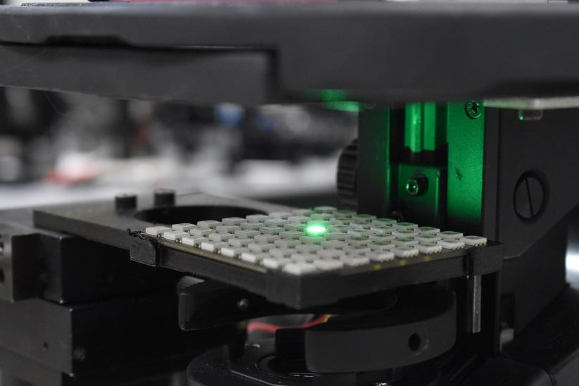
The course consists of four technology demonstrators (the last of technologies was included in the May course edition): digital holographic microscope, Fourier Ptychographic microscope, optical diffraction tomograph, and lensless holography system. The course aims to familiarise the widest possible group of people with these devices. During the one-day course, tutors train the course participants in the design, construction, calibration, and validation of each of the presented systems. The course consists of theoretical and practical parts.
"We strive to ensure that our techniques support, but also compete with fluorescent imaging methods," says Arkadiusz Kuś, PhD. "The proposed techniques are constantly developing along with the development of computing powers, thanks to which the study of biological objects is largely carried out not in equipment, under a microscope, but on a computer, on the computational side. This allows us to observe the impact of drugs or phototherapy in real time,” he explains.
The techniques used by PW scientists allow us both to view a given cell and what is happening to it, but also to accurately measure the cell and verify the correctness of the calculations made thanks to the created phantoms.
"We use the Nanoscribe Photonic Professional GT2 3D printer at the Warsaw University of Technology, which allows us to produce three-dimensional objects on a very high-resolution scale, corresponding to the scale at which the smallest elements of cells operate," says Michał Ziemczonok, MSc. "The printer uses polymer, which facilitates manoeuvring the degree of curing the prints, and this allows you to change the refractive index of the structure. As a result, we obtain objects that can be used optically and geometrically as cell models or other biological structures," he explains.
PW specialists hope that in the future their techniques will support histopathological diagnostics, which is extremely time-consuming, and the number of samples delivered to laboratories is constantly growing.
"Using our techniques, it is possible to perform a preliminary screening analysis of cells, without staining. This would facilitate faster work, because only those cells that have suspicious features would be subjected to closer examination, instead of all of them, as until now," explains Piotr Zdańkowski, PhD.
Future plans
The researchers are already making plans for the future, wishing to continue the project in the form of an Excellence-Center.
"These are three-day courses, extended with new technologies, allowing for a better exploration of phase imaging techniques," says Professor Małgorzata Kujawińska.
-
The project "qPI3Cells: Quantitative phase imaging at the cellular level" is financed by the POB Photonic Technologies of the Excellence Initiative - Research University programme, which is realised at the Warsaw University of Technology.
Team members:
Anna Pakuła, PhD; Arkadiusz Kuś, PhD; Piotr Zdańkowski, PhD; Maciej Trusiak, PhD; Mikołaj Rogalski, MSc; Michał Ziemczonok, MSc; Piotr Arcab, MSc; Professor Małgorzata Kujawińska; Michał Ziemczonok, MSc.


